
Guiding Images
If clarity and compassion matter...choose JRMC.
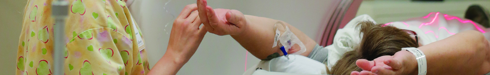
The radiology department is a vital part of Jamestown Regional Medical Center. What began in 1929 as two x-ray rooms and a darkroom, transitioned to an expansive multi-modality area with state-of-the-art technology. JRMC Radiology performs more than 16,000 exams each year.
JRMC Radiology is entirely digital; all images are produced, displayed, interpreted and archived using Picture Archiving and Communication System (PACS). With proper security, this allows providers to view images on monitors anywhere in or outside the building.
accredited


comprehensive
Let your doctor know you prefer your care at JRMC. Click on the technology below to learn more or download prep tips for your procedure.
The JRMC Radiology team performs x-rays of the chest, abdomen, spine, extremities and other bone or soft tissue areas of the body. Images are immediately available for viewing by the radiologist or ordering physician.
Our in-house MRI unit is the latest-generation magnetic resonance imaging scanner that scans quicker, reducing exam time. The system is less intimidating and less claustrophobia inducing than other MRI machines, contributing to patient comfort.
MRI stands for Magnetic Resonance Imaging. Without using traditional x-ray imaging, MRI allows doctors to see inside the body to diagnose and identify possible medical conditions. An MRI simply samples signals from the water that makes up your body. Specialized antennae create highly defined images that can be used to make diagnoses.
You’ll discover that MRI testing is painless and much quicker than you think. In fact, you’ll probably be very comfortable as you lie on the padded table. The accuracy and speed of the latest MRI scanners mean that you will be done quickly and your doctor will have to run fewer if any, follow-up scans.
CT (computerized tomography) scan of the body is a special x-ray examination that produces a series of cross-sectional pictures or images of the body. During the procedure, the x-ray tube moves in a large circle as it scans or rotates around your body. Receptors relay information to a computer that converts the data into images on a video screen. A radiologist will study these images and give a report to your physician. CT scans can distinguish bone, tissue, fat, gas, fluid, etc.
They are used to evaluate organ size and shape, evaluate masses, and diagnose pathology such as tumors cysts, aneurysm, ascites, lymphadenopathy, metastasis, ruptured discs, etc. CT scans can be performed with or without IV contrast.
JRMC was the FIRST facility in North Dakota to offer 3D mammography for breast cancer screening. 3D mammography (breast tomosynthesis) helps radiologists identify and characterize individual breast structures without the confusion of overlapping tissue.
Click the ribbon to learn more about how JRMC is removing all obstacles to receiving proper breast screenings.
JRMC has a primary ultrasound suite with a dedicated private bathroom, changing area and waiting room. It has been designed to provide space for caregivers and family members who accompany patients for an ultrasound procedure, especially the families of Family BirthPlace patients.
Ultrasound is the use of sound waves to obtain a medical image or picture of various organs, tissues or blood vessels in the body. Ultrasound procedures performed at JRMC include scans of the heart, abdomen and OB/GYN. These include small areas such as breast, thyroid and testicle and vascular scans.
Nuclear medicine imaging utilizes radiotracers, special cameras, and a computer to produce images of the interior of your body and allow staff to diagnose and determine the presence of disease based on its molecular structure.
Primary procedures performed in nuclear medicine include bone, gall bladder and thyroid uptake scans and rest/stress scans of the heart.
Bone Densitometry is a radiology procedure that determines bone mineral density or bone mass. A dedicated dual-energy x-ray absorptiometry (DEXA) unit will be used to produce x-ray images of the spine and hip. The unit will generate a computerized report that will include an image of the tested area, the bone mineral density measurement, and the T-score, which is a comparison to the peak bone mass measurements in a young population of the same gender and race.
Bone mass measurement studies provide information that will determine your risk of fractures and/or osteoporosis. If you are diagnosed with osteoporosis, your doctor may start a treatment plan to slow down or stop bone loss and reduce your risk of fracture.
During a comprehensive examination with DXA, you will lay comfortably on a padded table while the DXA unit scans two or more areas, usually the fracture-prone hip and spine. Unlike typical x-ray machines, radiation exposure during bone densitometry is extremely low. The entire process takes only minutes to complete. It involves no injections or invasive procedures.
Advanced Body Composition assessment produces images displaying the distribution of fat, lean tissue and bone translating the information into an easy to interpret the report. One of the key factors is Fat Mass index (FMI) an obesity classification scheme which measures the ratio of fat mass to height squared. Just like Body Mass Index (BMI), FMI is expressed in units of kg/m. FMI may be a better indicator of obesity than BMI because FMI is based on fat mass, not body weight, which is composed of both fat and lean constituents.
The body composition is a quick three to five-minute scan. Patients receive a low dose x-ray while lying on a comfortable padded table. There are no injections and it is non-invasive and painless. After the scan patients receive a detailed color report to give to their referring physician.
An echocardiogram evaluates the size of the chambers of the heart, including the dimension or volume of the cavity and the thickness of the chamber walls. Echocardiography can identify if the heart is pumping poorly or if one or more isolated areas have reduced movement. The pumping power of the heart will be calculated as the EF (ejection fraction). Echocardiography identifies the structures, thickness, and movement of each heart valve. It can evaluate valves for scarring, calcification, leaking and narrowing.
For patients in heart failure, an echocardiogram can assess the blood volume that returns from the lower part of the body. Electrocardiograms can also evaluate for congenital heart disease, blood clots or tumors in the heart, infection of heart valves, and abnormal elevation of pressure in the lungs. Echocardiography is useful in the diagnosis of fluid in the pericardium (the sac that surrounds the heart) and can determine when it is severe and potentially life-threatening.
Fluoroscopy produces a real-time moving image of the internal structures of a patient through the use of a fluoroscope. It uses digital detectors for timely acquisition and improved display of images.
Swelling in the bones, muscles, tendons, joints, ligaments, cartilage and other tissues are a common source of pain. Image-guided injection of a short-acting anesthetic and anti-inflammatory steroid is useful in the management of joint pain that has not resolved after conservative therapy. The joint injections can be used to:
- resolve pain faster than would occur with rest and medication
- delay surgical intervention
- diagnose the source of pain
- diminish symptoms
- control pain in non-surgical patients
Segmental pressures is a procedure performed to identify narrowing or blockages in the arteries in the lower legs. It is performed in patients who have pain in their legs, particularly pain with walking or exertion.
Always ready
JRMC Radiology is open 24/7. A board-certified radiologist is on site daily and is available for readings evenings, weekends and holidays.
There is no need for patients to travel for quality testing. Ask your doctor to refer you to in-town care at JRMC.